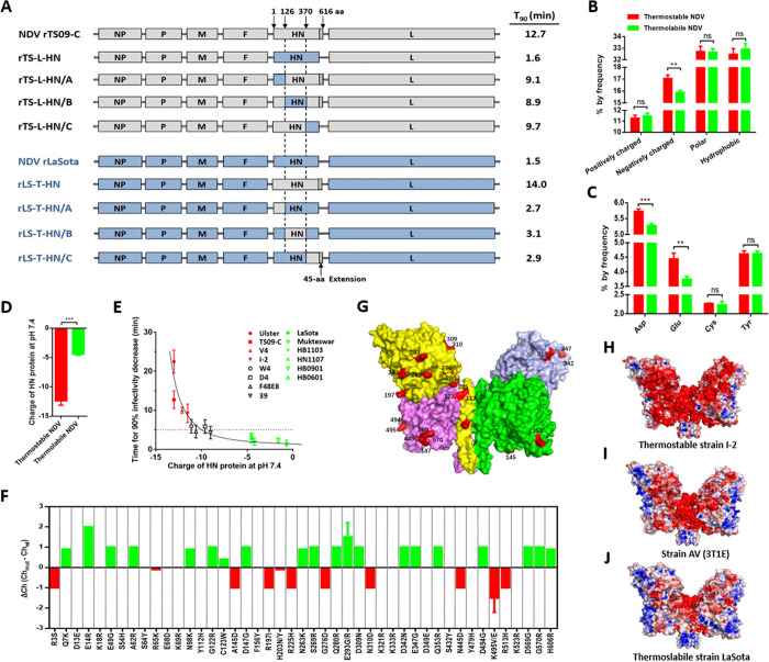
Fig 1. Charge of HN protein and its relationship with NDV thermostability.
(A) Schematic representation showing the construction of chimeric NDVs. Grey and blue bars represent the genes of TS09-C and LaSota strain, respectively. Corresponding nucleotide numbers where the HN fragments are fused using In-fusion cloning technology are depicted. For each chimeric virus, the viral thermostability (Time for 90% infectivity loss, min) at 56°C is indicated at right side of the diagram. (B) Percentage of amino acids with different characteristic in HN proteins from thermostable and thermolabile NDV strains. (C) Negatively charged amino acids composition of HN proteins from thermostable and thermolabile NDV strains. (D) Theoretical charge of HN proteins from thermostable and thermolabile NDV strains. The four thermostable strains are TS09-C, V4, I-2, and Ulster. The four thermolabile strains are LaSota, Mukteswar, HB1103, and HN1007. The theoretical charge of protein at pH 7.4 is determined by using IPC software (http://isoelectric.ovh.org), based on its amino acid sequence. (E) Scatter diagram showing the relationship between charge of HN protein and thermostability of NDV at 56°C. Each point represents the data from one NDV isolate. (F) Effect of each charged amino acid substitution on the charge of HN protein of NDV strain TS09-C. (G) Mapping of charge-associated amino acid substitutions onto the surface of HN protein of NDV strain TS09-C. The 23 charge-associated amino acid substitutions are labeled on the protein surface and colored red. (H-J) Molecular surfaces of thermostable and thermolabile NDV HN proteins are colored according to electrostatic potentials with a range of red (- 5.0 V) to blue (+ 5.0 V). The structures of HN proteins from NDVs are obtained by homology modeling, using the HN structure of AV strain (PBD ID code 3T1E) as a template.

