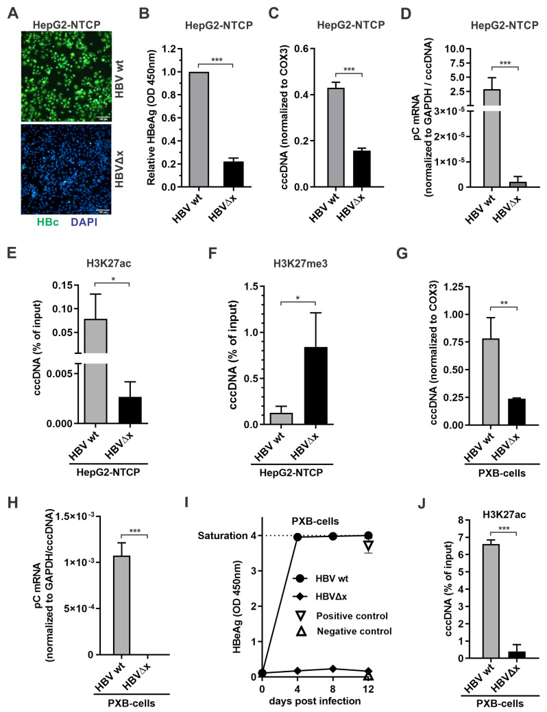
Fig 4. Characterization of the dependency of cccDNA transcription on HBx during HBV infection.
(A-F) HepG2-NTCP cells were infected with wt HBV and HBVΔx viral particles at 500 vge/cell for 10 days, and the following assays were performed: A) Intracellular HBV core protein (HBc) was detected by immunofluorescence, cell nuclei were stained by DAPI. Scale bar = 100 μm; B) Supernatant HBeAg was measured by ELISA (OD 450nm values; mean ± SD, n = 3); C) Levels of cccDNA were detected by qPCR and normalized to COX3 (mean ± SEM, n = 3); D) The relative levels of intracellular HBV pC mRNA was detected by qPCR and normalized to GAPDH and cccDNA (mean ± SEM, n = 3); E) The cccDNA-associated histone PTM H3K27ac was assayed by ChIP-qPCR and shown in percentage (%) of input (ΔCq; mean ± SEM, n = 3); F) The cccDNA-associated histone PTM H3K27me3 was analyzed by ChIP-qPCR and shown in percentage (%) of input (mean ± SEM, n = 3). (G-J) PXB-cells cells were infected with HBV wt and HBVΔx viral particles at 500 vge/cell for 12 days. G) Levels of cccDNA were detected by qPCR and normalized to COX3 (mean ± SEM, n = 3); H) HBV pC mRNA and I) HBeAg were analyzed as described above; J) cccDNA-associated H3K27ac was analyzed by ChIP-qPCR and shown in percentage (%) of input (mean ± SEM, n = 3). *p<0.05, **p<0.01, ***p<0.001.

