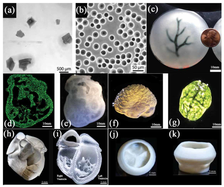
Figure 5.
(a) Gelatin particles prepared by mechanical force. (b) Gelatin microspheres prepared by coacervation method. (c) A schematic diagram of the arterial tree printed in a gelatin microgel supporting bath. (d) A cross section of the 3D printed heart in fluorescent alginate. (e) Dark field image of the 3D printed heart. (f) Side view of the brain printed with alginate. (g) The top view of the 3D printed brain. (h) MRI-derived 3D human heart scaled to neonatal size. (i) FRESH-printed collagen heart. (j,k) Top and side views of the FRESH-printed collagen heart valve with barium sulfate added for X-ray contrast. Scale bars: 10 mm in (c) and 1 cm in (d–g); (a,c,d,f,g) are from reference [45], Reprinted with permission from Ref. [45]. Copyright 2015, copyright Hinton et al. (b,h,i,j,k) are from reference [38], Reprinted with permission from Ref. [38]. Copyright 2019, copyright Lee et al.

