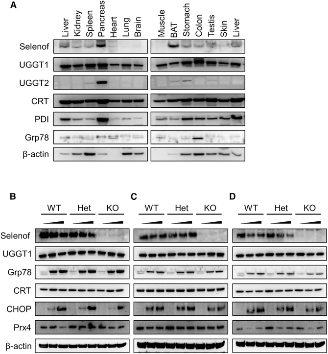
Figure 4. Selenof Deficiency Does Not Lead to ER Stress.
(A) Expression pattern of Selenof and other relevant proteins across mouse tissues.
(B-D) Expression of Selenof and other relevant proteins in MEFs from WT, heterozygous, and Selenof KO mice subjected to ER stressors thapsigargin (B), tunicamycin (C), and brefeldin A (D). WT, heterozygous, and KO MEFs were treated with two concentrations of stressors along with control (DMSO treated): thapsigargin (5 nM and 50 nM), tunicamycin (50 ng/mL and 500 ng/mL), and brefeldin A (0.5 μM and 5 mM). Proteins assayed are shown on the left.

