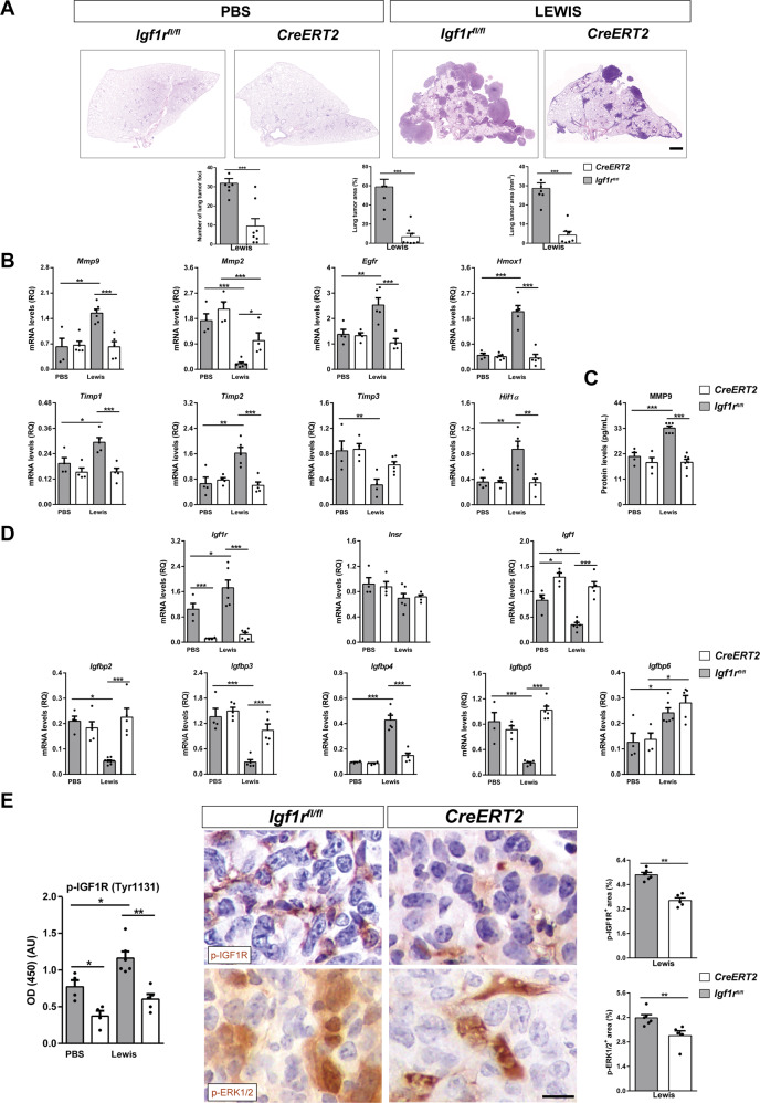
Fig. 4. Reduced tumor burden and decreased expression of metastasis markers, p-IGF1R and p-ERK1/2, as well as changes in IGF system gene expression in lungs of IGF1R-deficient mice.
A Representative histopathology images of lung metastasis (H&E) and respective quantifications of lung foci and lung tumor area (% and mm2) in IGF1R-deficient (CreERT2) vs. Igf1rfl/f mice (n = 5–8 mice per group; Scale bar: 100 µm). B Lung tissue mRNA expression levels of Mmp9, Mmp2, Egfr and Hmox1 (tumor progression), Timp2 and Timp3 (inhibitors of metalloproteinases), and Hif1α (hypoxia) markers, normalized to 18 S expression in PBS- or LLC-challenged, and C MMP9 protein levels in lung homogenates of CreERT2 vs. Igf1rfl/fl mice (n = 4–7 mice per group). D Lung tissue mRNA expression of IGF system-related genes Igf1r, Insr, Igf1, Igfbp2, Igfbp3, Igfbp4, Igfbp5 and Igfbp6 normalized to 18 S expression in PBS- or LLC-challenged CreERT2 vs. Igf1rfl/f mice (n = 5–7 mice per group). E p-IGF1R protein levels in lung homogenates, as well as representative immunostains for p-IGF1R and p-ERK1/2 (p-42/44) and respective quantifications of p-IGF1R+ and p-ERK1/2+ areas (%) (brown) in lung metastatic tumors of PBS- or LLC-challenged CreERT2 vs. Igf1rfl/fl mice (n = 4–6 mice per group; Scale bar: 15 µm). Quantifications were performed randomly in five different fields. Data are expressed as mean ± SEM. *p < 0.05; **p < 0.01; ***p < 0.001 (Mann-Whitney U test or Student´s t-test for comparing two groups and the Dunn-Sidak test for multiple comparisons).

