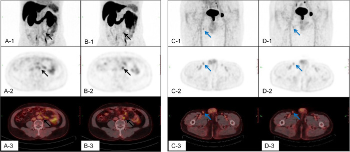
Fig. 1. The 18F-DCFPyL uptake of malignant and benign lymph nodes in dual-phase PET/CT imaging.
The A,B,C, D-1 were the maximum intensity projection (MIP) images; The A,B,C, D-2 were the cross sections of PET; and the A,B,C, D-3 were the cross sections of fusion images. The A, C-1, 2, 3 were for the early phase, and the B, D-1, 2, 3 were for delay phase. The A and B were the dual-phase 18F-DCFPyL PET/CT scan for a retroperitoneal lymph node metastasis. The short diameter of this lymph node was about 7 mm, SUVmax early was 3.0, SUVmax delay was 3.8, △SUVmax was 0.8, RI was 26.7%, SUV ratio early was 5.0, SUV ratio delay was 12.7, and ratio(SUV ratio delay/early) was 2.54. The C and D were the dual-phase 18F-DCFPyL PET/CT scan for a reactive hyperplastic lymph node. The short diameter of this lymph node was about 7 mm, SUVmax early was 2.1, SUVmax delay was 1.7, △SUVmax was -0.4, RI was -19.0%, SUV ratio early was 3.5, SUV ratio delay was 3.4, and ratio (SUV ratio delay/early) was 0.97.

