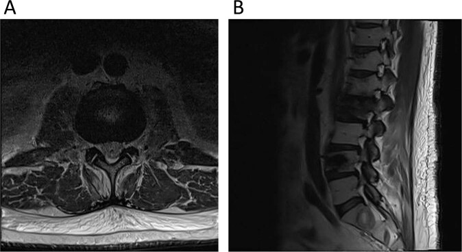
Fig. 1. Non-contrast MRI of the lumbar spine.
A Axial image at the L2-L3 level demonstrating bilateral neuroforaminal stenosis. B T2-Weighted Images MRI Images of the lumbar spine demonstrating pathologic compression fractures of the L2 and L4 vertebral bodies along with multilevel stenosis of the right neuroforamen.

