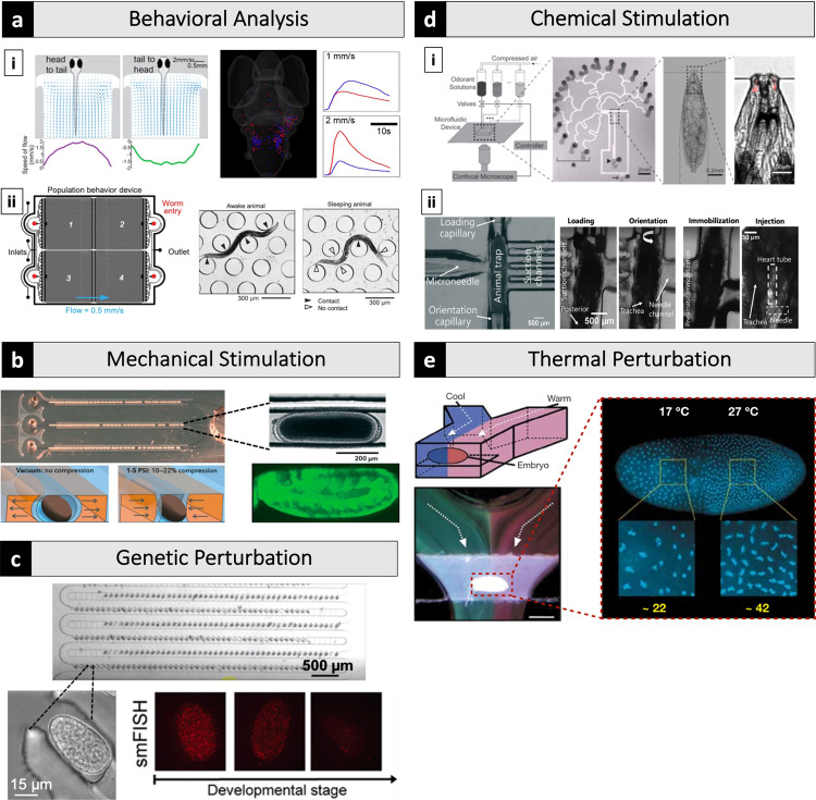
Fig. 3. Microfluidic systems can deliver stimulations with high precision and at throughputs not previously attainable while making once laborious experiments less demanding.
a-i A 3D-printed microfluidic device compatible with a custom-built light-sheet microscope to stimulate zebrafish with precise flow vectors for brain-wide calcium imaging40. a-ii A microfluidic device with an array of microposts for analysis of sleep behavior of C. elegans46. b A microfluidic system that automatically aligned Drosophila embryos and precisely compressed them using pneumatically actuated deformable sidewalls with simultaneous live imaging57. c A microfluidic system capable of trapping hundreds of C. elegans embryos quickly and enabling efficient reagent exchange59. d-i A microfluidic system visualized the response of olfactory receptor neurons (ORNs) of Drosophila larva in response to controlled odorant exposure69. d-ii A microfluidic device with integrated glass capillaries and a microneedle for chemical injection of Drosophila larvae70. e A microfluidic device exposed Drosophila embryo to a thermal gradient along the anterior-posterior axis using two laminar flow streams with different temperatures76. All panels are cropped and adapted versions of the originals. Panel c is adapted with permission from Charles et al.59, copyright 2020 American Chemical Society.

