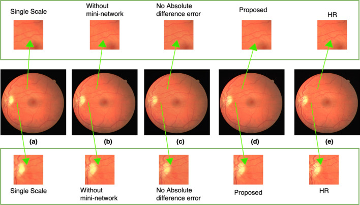
Figure 16.
Results for ablation studies. The upper and lower rows show the extracted small region. (a) Represents the resultant SR image with single scale features extraction. (b) Represents the resultant SR image for experiment without the mini-network. (c) Represents the resultant SR image for experiments after removing the absolute difference error term from the loss function. (d) Represents the resultant SR image for experiment with the overall proposed method. (e) Represents the corresponding HR ground truth image. Arrows point to the extracted small regions to highlight the regions where difference occurs.

