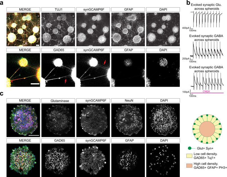
Fig. 3. Characterizing the cellular architecture of MoNNet.
a Whole-mount immunostaining of four weeks old MoNNets. Top row: maxima projections showing Tuj1, Syn-GCaMP6f, GFAP and DAPI labeling. Bottom row: a confocal optical section showing co- or exclusive (red arrow) labeling of interconnections with GAD65 or synGCaMP6f signals, suggesting existence of excitatory as well as inhibitory signaling transmission across spheroids unit in MoNNets. Scale bars are 100 μm. These representative experiments were independently repeated >3 times. b Electrophysiology recordings validating the existence of monosynaptic functional glutamatergic and GABAergic signal transmission across spheroids. For all recordings, a neighboring spheroid, located ~250–300 μm, away was stimulated at 10 Hz. Top-to-bottom: representative 1 s trace of a cell held at −70 mV showing robust evoked excitatory synaptic transmission across spheroids; representative 1 s trace of a cell held at 0 mV showing robust evoked inhibitory synaptic transmission across spheroids; representative 1 s trace of a cell held at 0 mV showing robust evoked inhibitory synaptic transmission during CNQX (10 μM) blockade of glutamatergic transmission. c, Immunostaining of 2 weeks old individual spheroids vibratome sections (50 μm thick) to visualize their cellular architecture. Glutaminase and NeuN immunolabeling revealed preferentially peripheral localization of glutamatergic cells, whereas the GAD65 and GFAP expression is preferentially localized in the inner regions. A schematic summary is shown on right. Scale bars are 100 μm. These representative experiments were independently repeated >3 times. Also see Supplementary Fig. 7 for additional GAD65 stainings, Supplementary Fig. 8 for PH3 and activated Caspase3 labeling, and Supplementary Video 7 for Confocal z-stack showing the 3D cellular architecture of MoNNet.

