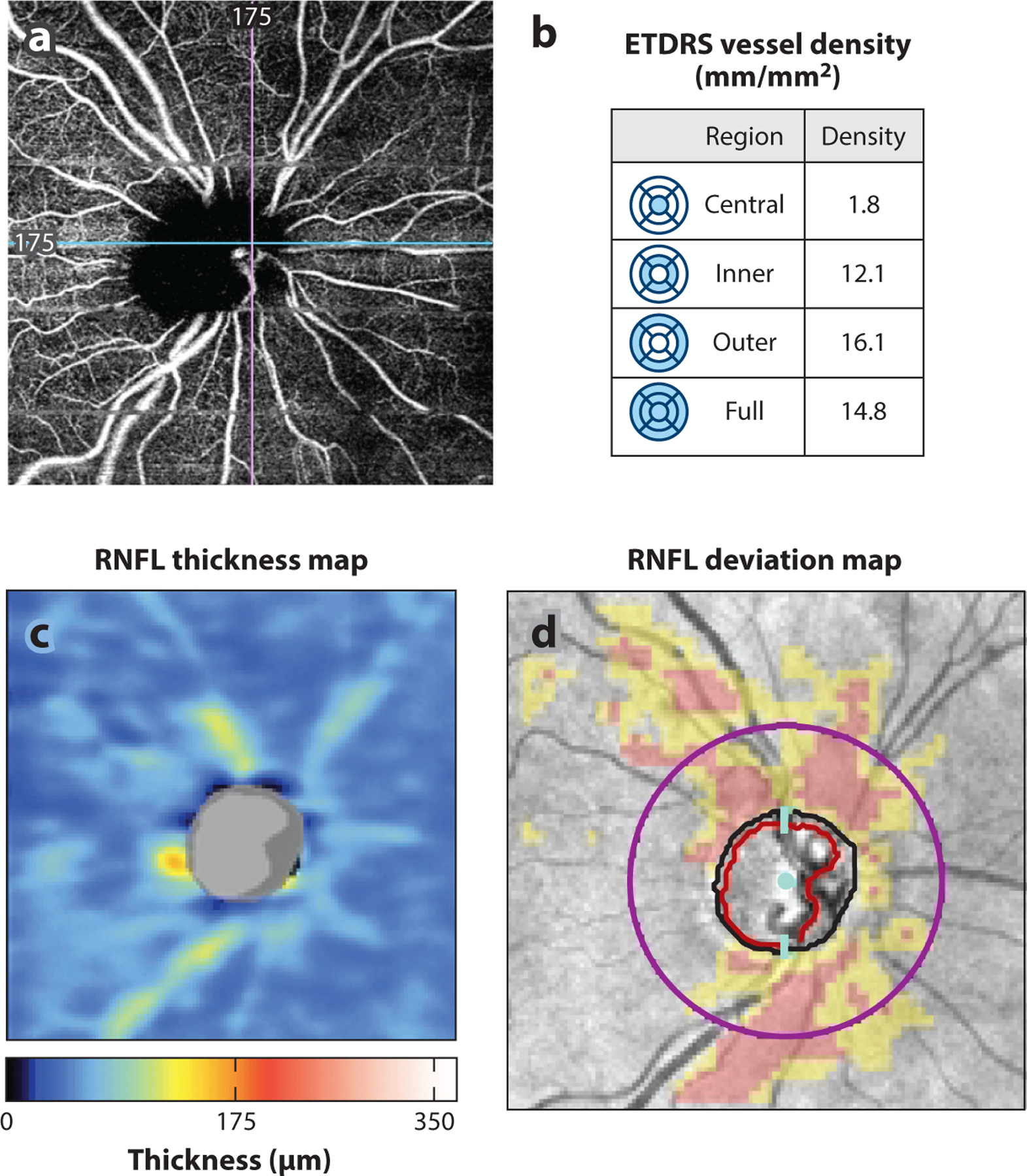
Figure 5.

(a) AngioPlex OCT-angiography (Carl Zeiss Meditec, Dublin, California) image of a glaucomatous eye. (b) Peripapillary superficial vessel density (mm/mm2) in the central, inner, outer, and full regions is quantified in the table. (c,d) Region of decreased vessel density correlates with region of RNFL thinning as seen on (c) the RNFL thickness map and (d) the RNFL deviation map from the Cirrus HD-OCT (Carl Zeiss Meditec) report. Abbreviations: ETDRS, Early Treatment Diabetic Retinopathy Study; HD-OCT, high-definition OCT; OCT, optical coherence tomography; RNFL, retinal nerve fiber layer.
