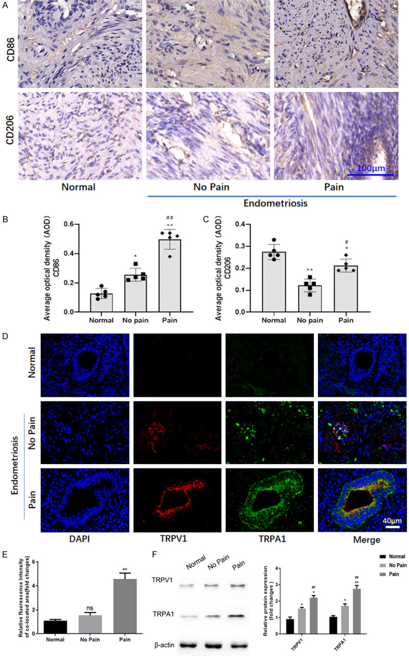
Figure 1.

Macrophage polarization and TRPV1/TRPA1 heteromers in human endometriosis tissues. A. The expression of CD86 and CD206 in endometriosis tissues was detected by immunohistochemistry. B. The expression of CD86 in endometriosis tissues detected by immunohistochemistry was indicated by average optical density (AOD). *, **denote P<0.05 and P<0.01, compared with Normal group, ##denotes P<0.01, compared with No pain group. n=5. C. The expression of CD206 in endometriosis tissues detected by immunohistochemistry was indicated by average optical density (AOD). *, **denote P<0.05 and P<0.01, compared with Normal group; #denotes P<0.05, compared with No pain group. n=5. D. The fluorescent expression of TRPV1 and TRPA1 in human endometriosis tissues. E. The quantification of immunofluorescence for co-expression of TRPV1 and TRPA1. n=3. Ns, no significant difference. **P<0.01, compared with No pain group. F. The protein expression of TRPV1 and TRPA1 in human endometriosis tissues. *, **denote P<0.05 and P<0.01, compared with Normal group; #, ##denote P<0.05 and P<0.01, compared with No pain group. n=3.
