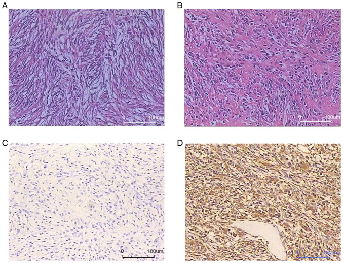
Figure 3.
Hematoxylin and eosin staining, and immunohistochemistry. (A) Fibroblastic spindle cells and inflammatory infiltrate upon hematoxylin and eosin staining (magnification, ×200). (B) Mucinous degeneration identified by hematoxylin and eosin staining (magnification, ×200). (C) ALK detection by immunohistochemistry, showing a lack of staining (magnification, ×200). (D) Positive vimentin detection by immunohistochemistry (magnification, ×200).

