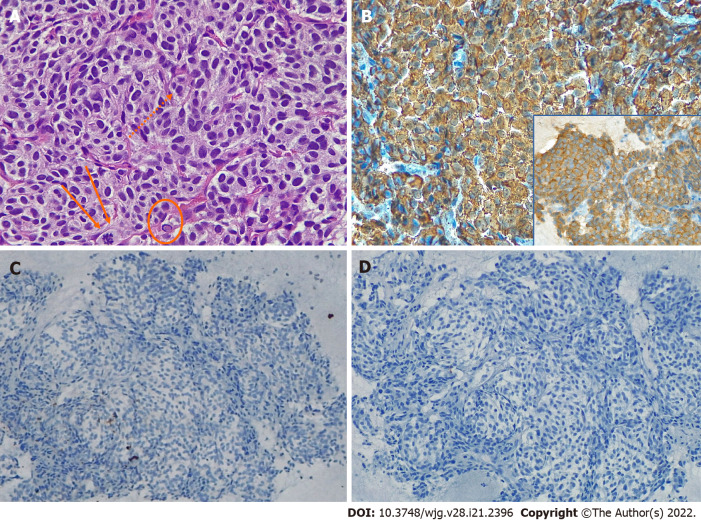
Figure 2.
Fine needle biopsy findings. A: Proliferation of small to medium-sized cells arranged in a nest pattern was evident, the cells occasionally showed small nucleoli (dots), mitotic figures (arrows), and intra-cytoplasmic hyaline globules (encircled), hematoxylin and eosin original magnification (O.M) × 40; B: Chromogranin A (CgA) positivity of neoplastic cells; inset: Synaptophysin positivity of neoplastic cells, CgA stain, O.M. × 20; B inset, synaptophysin stain, O.M. × 20; C: AE1/AE3 cytokeratins expression in scattered cells, cytokeratin AE1/AE3 stain, O.M. × 20; D: S100 negativity, S100 stain, O.M. × 20).

