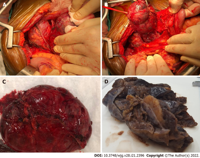
Figure 3.
Macroscopic findings. A and B: At surgical exploration a large well-defined cystic mass located underneath the mesocolon plane was found; C: Radical enucleation of the lesion; D: Grossly, the cystic lesion showed a thick fibrous wall with a solid component and a yellowish, lobulated appearance on cut surface.

