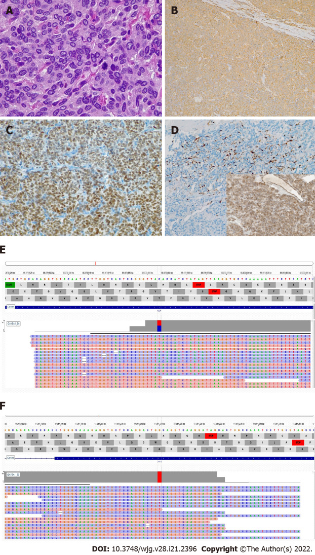
Figure 4.
Histologic and molecular findings. A: The lesion was composed of nests and cords of polygonal cells with abundant granular cytoplasm, and occasional mitotic figures in the more cellular areas, hematoxylin and eosin original magnification (O.M) × 60; B: Chromogranin A (CgA) positivity in neoplastic cells; inset: Synaptophysin positivity in neoplastic cells, CgA stain, O.M × 10; C: GATA-3 positivity in neoplastic cells, GATA-3 stain, O.M. × 10; D: S100-positive cells were scattered; inset: Succinate dehydrogenase subunit B (SDHB) expression was preserved, S100 stain, O.M. × 10; inset, SDHB stain, O.M. × 10; E: KDR mutation; F: JAK3 mutation.

