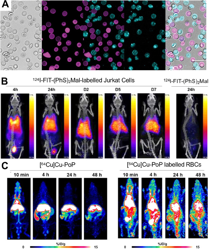
Figure 11.
(A) Microscopy images of RBCs labeled with either Cy3 or Cy5 dyes based on [18F]BF3-Cy3-NHS. Bright field imaging of the RBC-Cy3/RBC-Cy5 mixture (far left). Middle left image: RBC-Cy3s and middle right image is for RBC-Cy5. Middle image is an overlay of the RBC-Cy3 and RBC-Cy5 showing a lack of spectral overlap between the two fluorophores, and no mixing of fluorophores between cells after 14 h. Far left image is an overlay of bright field and fluorescent images. Adapted with permission from Wang et al., ref (153). Copyright 2017 SAGE Journals. (B) PET/CT images of NSG mice that received 124I-FIT-(PhS)2Mal labeled Jurkat cells at 4 and 24 h and 2, 5, and 7 days or 124I-FIT-(PhS)2Mal at 24 h post IV injection. Adapted with permission from Pham et al., ref (156). Copyright 2020 American Chemical Society. (C) PET image of mice injected with 64Cu-labeled porphyrin-phospholipid conjugate (PoP) (left) or 64Cu-labeled PoP RBCs (right). RBCs were obtained from mice prior to labeling and intravenous injection. Adapted with permission from Kumar et al., ref (162). Copyright 2021 Kumar et al. Published by Wiley-VCH GmbH under CC License [https://creativecommons.org/licenses/by/4.0/].

