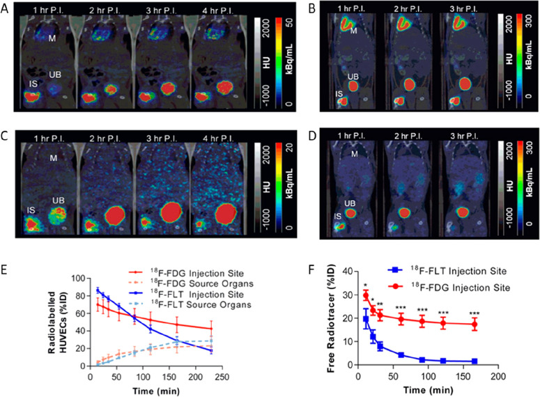
Figure 14.
Comparison of human vascular endothelial cells (HUVECs) labeled with [18F]FDG (A) and [18F]FLT (C) or free [18F]FDG (B) and [18F]FLT (D) injected at the same site, representative averaged (1 h) images from each hour postinjection (P.I.). IS: Injection site. M: Myocardium. UB: Urinary bladder. (E, F) Time-activity curves for 18F-labeled cells and the corresponding radiotracers. The persisting signal at the injection site with [18F]FDG-labeled cells is partly due to [18F]FDG leaking from the labeled cells and being taken up by neighboring tissue. Note the increasing 18F signal in the heart region, indicating release of [18F]FDG into the circulation. In contrast, free [18F]FLT is rapidly cleared from neighboring tissue. Consequently, the signal from [18F]FLT-labeled cells is more representative of the presence of labeled cells at the injection site. Adapted with permission from Macaskill et al., ref (180). Copyright 2017 Springer Nature under CC License [https://creativecommons.org/licenses/by/4.0/].

