
Figure 18.
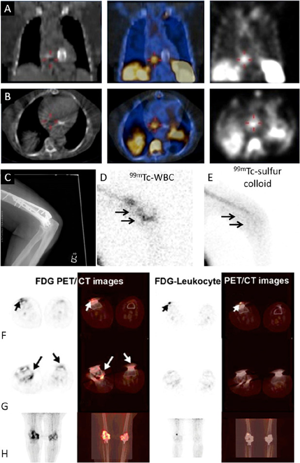
Infection and inflammation imaging with radiolabeled WBC. (A, B) [99mTc]Tc-HMPAO-WBC SPECT/CT images of a patient with endocarditis of native tricuspid valve: (A) coronal views and (B) transaxial views; CT (left), fused SPECT/CT (center), and SPECT (right). SPECT/CT allowed the exclusion of an initially suspected prosthesis-associated endocarditis. Adapted with permission from Erba et al., ref (341). Copyright 2012 SNMMI. (C–E) CT and scintigraphy images taken after administration of [99mTc]Tc-HMPAO-WBC (D) or 99mTc-sulfur colloid (E) showing prosthetic joint infection in the distal right humerus. Note the focal accumulation of radiolabeled WBC compared to the more diffuse pattern of the colloid. Adapted with permission from Palestro, ref (311). Copyright 2016 SNMMI. (F–H) PET/CT images of a patient who had undergone bilateral knee arthroplasty 1 year previously and presenting bilateral knee pain. Selected axial (F, G) and maximum intensity projection (H) PET/CT images are shown. The [18F]FDG PET/CT images (two left columns) show increased [18F]FDG uptake around the right knee prosthesis and slightly increased [18F]FDG accumulation around the left knee prosthesis. [18F]FDG-labeled WBC PET/CT images (two right columns) show intense WBC accumulation in soft tissue in the anterior part of right knee. The final microbiological diagnosis confirmed infection of the right knee prosthesis. The clinical diagnosis confirmed aseptic loosening of left knee prosthesis. Adapted with permission from Aksoy et al., ref (351). Copyright 2014 Springer Nature.