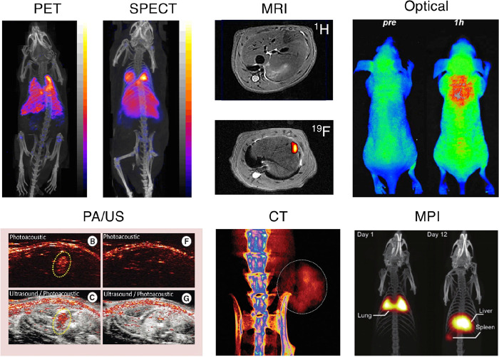
Figure 2.
Representative images showing preclinical cell tracking studies with different imaging modalities and cell types, including nuclear imaging-based techniques with 89Zr- and 111In-labeled 5T33 cells (PET and SPECT) Reproduced with permission from ref (3). Copyright 2015, Springer Nature under CC License [https://creativecommons.org/licenses/by/4.0/]. MRI with SPIO- and 19F-labeled mesenchymal stromal cells. Reproduced with permission from ref (4). Copyright 2020, Springer Nature under CC License [https://creativecommons.org/licenses/by/4.0/]. Optical cell tracking of human hematopoietic cells. Reproduced with permission from ref (5). Copyright 2004, Springer Nature. Photoacoustic (PA) and ultrasound (US) cell tracking with gold nanoparticle-labeled cells. Reproduced with permission from ref (6). Copyright 2012, PLOS One under CC License [https://creativecommons.org/licenses/by/4.0/]. CT cell tracking of gold nanoparticle-labeled T cells. Reproduced with permission from ref (7). Copyright 2015, American Chemical Society. MPI cell tracking with SPIO labeled-stem cells. Reproduced with permission from ref (8). Copyright 2016, Springer Nature under CC License [https://creativecommons.org/licenses/by/4.0/].

