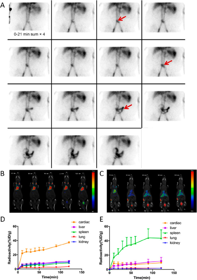
Figure 20.
Applications of radiolabeled RBCs. (A) Scintigraphic images of 99mTc-labeled RBCs showing bleeding originating from a branch of the superior mesenteric artery. A focus of increasing intensity is visible in the lower abdomen at the midline (arrows), showing anterograde and retrograde movement conforming to the bowel lumen. The focus crosses the midline several times and is therefore most compatible with small-bowel bleed. Adapted with permission from Grady. ref (325). Copyright 2016 SNMMI. (B, C) Coronal PET/CT images of untreated (B) and heat-stressed (C) [18F]FDG-labeled RBCs in mice, taken over 120 min. Untreated RBCs are mostly visible in the heart and carotid regions, with limited urinary excretion of [18F]FDG. In contrast, heat-stressed RBCs rapidly accumulate in the liver and spleen and increased release of 18F from the RBC is visible from the bladder signal. (D, E) Time-activity curves of 18F uptake in major organs after administration of untreated (D) and heat-stressed (E) [18F]FDG-labeled RBCs, mirroring the profiles observed on the PET/CT scans. Adapted with permission from Yin et al., ref (360). Copyright 2021 Springer Nature under CC license [https://creativecommons.org/licenses/by/4.0/].

