Figure 1.
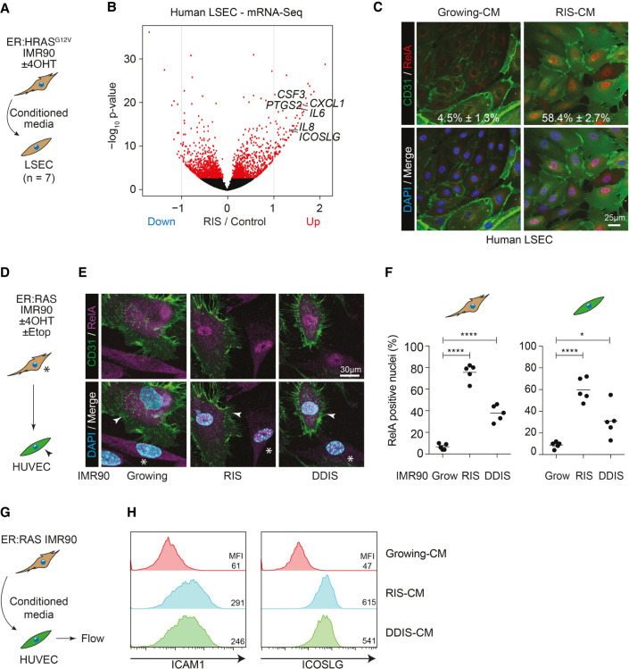
Senescent cells nonautonomously induce NF-κB activity in adjacent endothelial cells. (A) Experimental setup: Primary human liver sinusoidal endothelial cells (LSECs) were incubated in conditioned media (CM) from growing or RIS ER:HRASG12V IMR90 cells for 24 h before harvesting for mRNA sequencing. (B) Volcano plot of log2 fold change in gene expression against the −log10 P-value in RIS-CM compared with Grow-CM-treated LSECs. Red dots are significantly differentially expressed (DE; FDR < 0.05) genes. n = 7 biological replicates for all conditions. (C) Representative immunofluorescence of LSECs from a single donor treated with Grow-CM or RIS-CM and stained for CD31 and RELA. n = 3 biological replicates; mean ± SEM. Scale bar, 25 µm. (D) Experimental setup: direct coculture of growing, RIS, or DDIS (treated with etoposide) ER:HRASG12V IMR90 cells (asterisks) with HUVECs (arrowheads). (E) Representative immunofluorescence of coculture with senescence-dependent nuclear localization of RELA in both CD31− IMR90s and CD31+ HUVECs. Scale bar, 30 µm. (F) Separate quantification of RELA nuclear positivity from five biological replicates. Dots are individual replicates, and bars are means. Data were analyzed by one-way ANOVA with Sidak's multiple comparisons test; (*) P ≤ 0.05, (****) P ≤ 0.0001. (G) Experimental setup: HUVECs were incubated in CM from growing, RIS, or DDIS ER:HRASG12V IMR90 cells for 16 h before flow cytometry. (H) Representative flow cytometry histograms of ICAM1 (left) and ICOSLG (right) expression on HUVECs incubated in the indicated CM. n ≥ 3 biological replicates.

