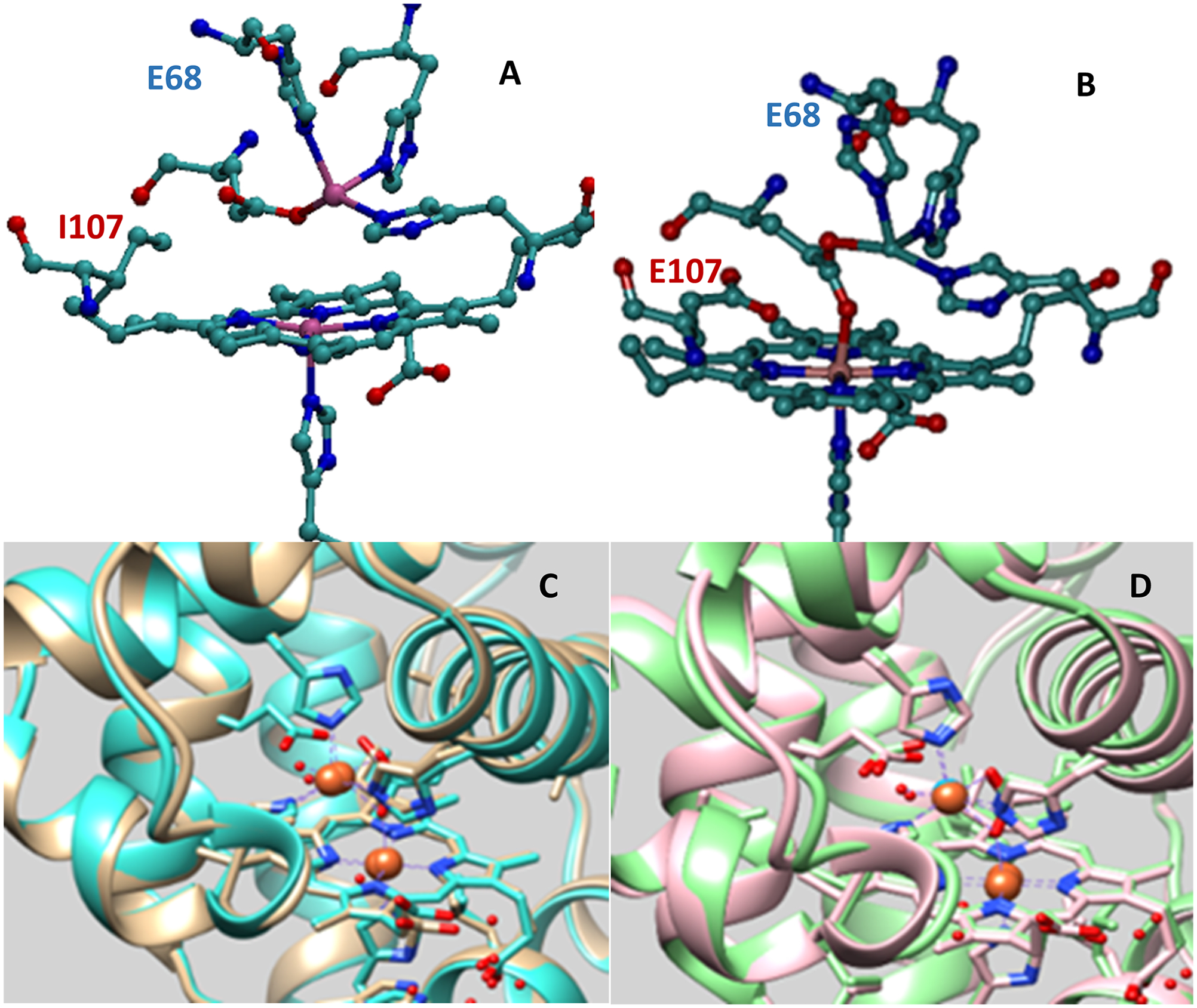
Figure 1.

Crystal structure of (A) FeB(II)-V68ECuBMb (PDB: 3K9Z, tan); (B) FeB(II)-V68E/I107ECuBMb (PDB: 3M39, turquoise); (C) overlay of the crystal structure of FeB(II)-V68ECuBMb (tan) and FeB(II)-V68E/I107ECuBMb (turquoise); and (D) overlay of the crystal structure of FeB(II)-V68E/I107ECuBMb (light green) and CuB(II)-V68E/I107ECuBMb (PDB: 3M3A, pink), water molecules, and Fe(II) are represented by red and brown, respectively.
