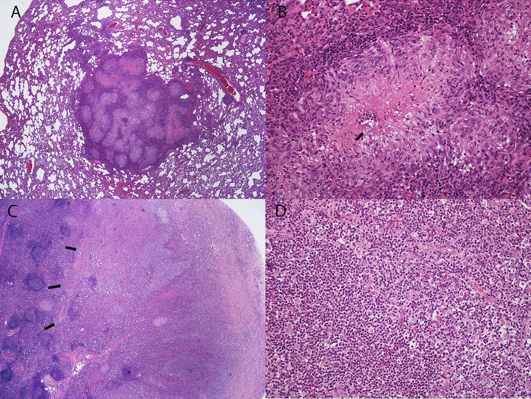
Fig. 5.

Examples of lung and mediastinal lymph node histopathology in Coccidioides-infected dogs from the Control group. (A) Multifocal to coalescing granulomatous and pyogranulomatous lesions with multifocal necrosis in lung lobe. (B) Spherule (arrow) in a necrotic center surrounded by pyogranulomatous inflammation. (C) There is effacement of normal architecture of the mediastinal lymph node due to severe pyogranulomatous inflammation (arrows). (D) Higher power image of lymph node effacement in (C) shows diffuse neutrophilic inflammation. Stain: hematoxylin and eosin. Magnification: A, C – 20X; B, D – 200X.
