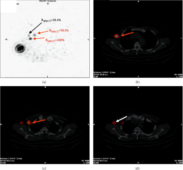
Figure 2.

A 60-year-old female patient with invasive ductal carcinoma of the right breast. (a) Maximum intensity projection images. Three hot nodes are visualized with different RSPECT. Two nodes with higher RSPECT (100%, 50.4%) are examined pathologically to be metastatic (red arrows), and another node with lower RSPECT (28.4%) is healthy (black arrow). (b–d), SPECT/CT hybrid images for localizing the two metastatic nodes (red arrows) and the healthy node (white arrow). The surgeon might selectively avoid excising lymph nodes with lower radioactivity (RSPECT ≤ 30%) deriving from preoperative SPECT/CT images.
