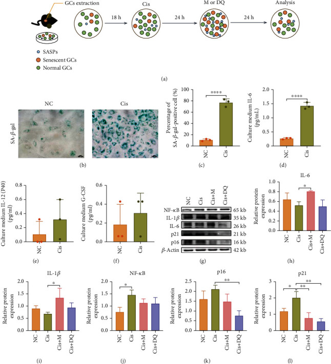
Figure 1.

Metformin and DQ independently reduced senescent granulosa cells and SASP caused by cisplatin in vitro. (a) Flowchart of primary GCs in vitro experiment. (b, c) Light microscopic images of SA-β-gal-stained GCs after cisplatin administration and quantitative analysis of SA-β-gal–positive GCs (n =3). Scar bars are marked on respective images. (d–f) Cytokines accumulated in cell culture medium of the NC and Cis groups. (g–l) The protein levels of senescence-associated markers and SASP were analyzed by western blotting (n =3). Results are presented using mean ± standard deviation values. ∗p < 0.05, ∗∗p < 0.01, ∗∗∗∗p < 0.0001.
