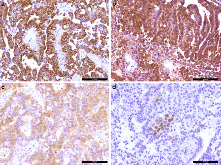
Fig. 5.
The tumors cells were positive in the immunohistochemical reaction for the pan-cytokeratin marker MNF116 (brown, a). The papilloma cells also show signal in the immunohistochemistry for transthyretin (prealbumin, b). Weak signal was observed in the immunohistochemical reaction for S100 (c). Tumor cells were partially labelled in the immunohistochemical reaction for cytokeratin 7 (brown, d). Hematoxylin (blue) was used as counterstaining in all cases (a–d). Scale bars: 100 µm

