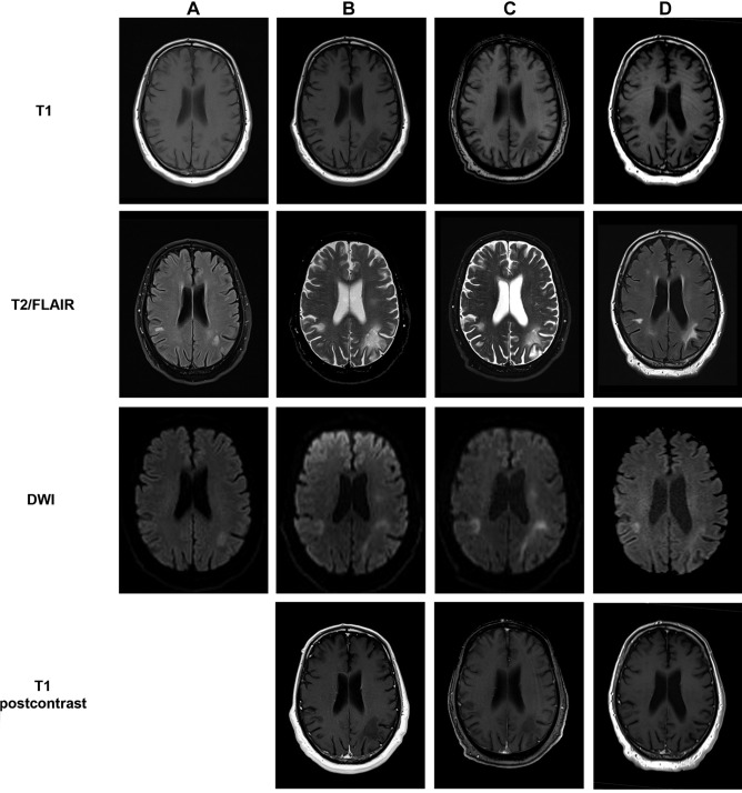
Fig. 1.
MRI of the head during treatment with pembrolizumab. Shown is a panel with sequential MRI scans of the head from July 2019 (A), before the third pembrolizumab infusion in January 2020 (B), the fifth pembrolizumab infusion in February 2020 (C), and after the final treatment with pembrolizumab in a follow-up examination in August 2020 (D). Shown sequences are T1 (A–D), T2-weighed (B–C) or FLAIR (A, D), DWI (A–D), and T1 postcontrast (B–C)

