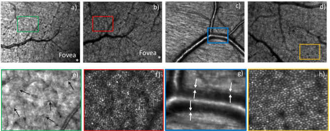
Figure 4.
AO-SLO images of a healthy volunteer recorded with a field of view of 4° × 3°. (a) AO-SLO image from near the fovea with its focus on the nerve fiber layer and (b) the same field, now focused on the photoreceptor layer. Images (c,d) are from two different fields, centered at ~ 4° eccentricity from the fovea and focused on the nerve fiber layer and the photoreceptor layer, respectively. Figures (e–h) are the 4 × enlarged view of the rectangular boxes marked in Figures (a–d), respectively. The arrows in (e) indicate small capillaries while in (g) the vessel wall is marked.

