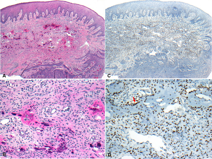
Fig. 1.
Photomicrographs showing a POF. A Photomicrograph showing a nodule of POF, covered by surface mucosa (H&E*, X 20). B The lesion is composed of a proliferation of spindled-shaped cells with bone formation (H&E*, X 200). C Photomicrograph showing the nodule of POF shown in A (IHC**, X 20) with nuclear staining of the lesional cells with SATB2 (D, IHC**, X 200), more prominently in those cells adjacent to the area of calcification (red arrow)

