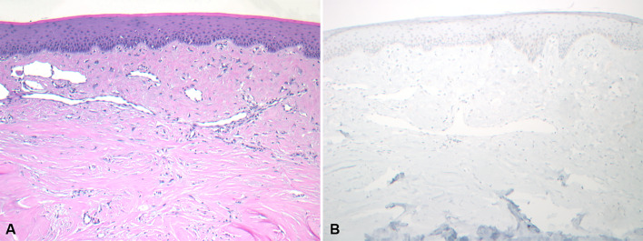
Fig. 3.
Photomicrographs showing a giant cell fibroma. A A giant cell fibroma characterized by large and stellate-shaped fibroblasts within the superficial connective tissue (H&E*, X 100) and B Negative SATB2 immunoreactivity in the lesional cells (IHC**, X 100). *Hematoxylin and eosin. **Immunohistochemistry

