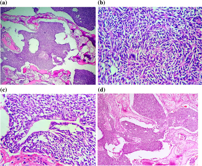
Fig. 4.
a A tumours composed of small epithelioid cells with hyperchromatic nuclei and minimal cytoplasms diffusely infiltrating in to bone. H&E × 4. b A photomicrograph showing epithelial whorls. H&E × 40. c Ameloblast like tall columnar cells with reverse polarity nuclei present throughout the tumour. H&E × 40. d A section showing areas reminiscent of Homer-Wright rosettes. H&E × 10

