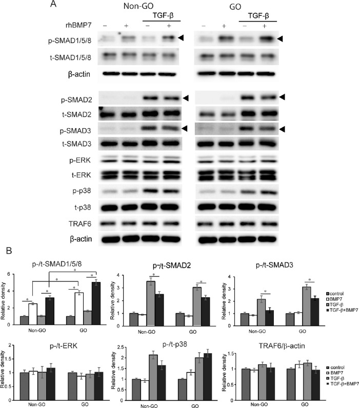
Figure 3.
Effect of BMP7 on the activation of signal proteins in response to TGF-β treatment. Orbital fibroblasts from GO patients (n = 3) and normal control non-GO subjects (n = 3) were treated with 5 ng/mL TGF-β for one hour with or without pretreatment with 100 ng/mL rhBMP7 for one hour. (A) In Western blot analyses, treatment of rhBMP7 resulted in a significant increase in the levels of phosphorylated forms of SMAD1/5/8 more predominantly in GO cells, while suppressed phosphorylation of SMAD2/3 was activated by TGF-β. Experiments were performed in three GO and three non-GO cells from different individuals. The representative gel images are shown. (B) Quantification of signal protein markers was performed using densitometry. The relative band intensity of each protein was indicated by the ratio of phosphorylated protein and total protein and normalized to the level of β-actin in the same sample. The results are presented as the mean density ratio ± SD (*P < 0.05).

