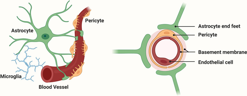
Fig. 1.
The cellular composition of the blood–brain barrier (BBB). The barrier is formed by a continuum of endothelial cells which line the interior of blood vessels (brown), and which are firmly attached to a basement membrane (BM) composed of a mix of extracellular matrix (ECM) proteins (pink). Pericytes (yellow) are located within the vascular BM. Astrocyte (green) endfeet contact the vascular BM, thus connecting blood vessels to neurons within the brain parenchyma. Microglia (blue) are also in close contact with astrocytes and blood vessels

