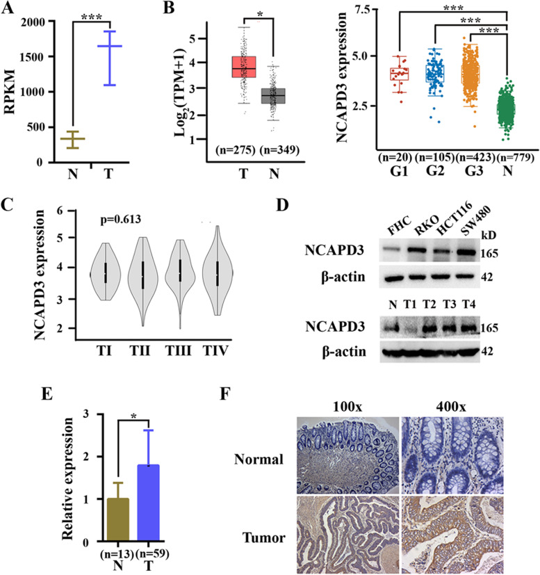
Fig. 1.
NCAPD3 was higher expression in human CRC. A Expressions of NCAPD3 in CRC tissues and corresponding non-tumor normal tissues of clinic specimen by analyzing our RNA-seq data. B The levels of NCAPD3 mRNA in CRC tissues and normal tissues were analyzed by using GEPIA (left) and Assistant for Clinical Bioinformatics (right) web tools. G1, G2, G3 represent different tumor grades. C The level of NCAPD3 at different tumor stages were conducted by using online database. D Protein levels of NCAPD3 in human normal colorectal mucosal cell line FHC, CRC cell lines (RKO, HCT116 and SW480) and clinic specimen (including CRC tissues and normal colorectal tissues) were detected by Western blot assay. E qRT-PCR analysis of mRNA expression in 59 human CRC tissues and 13 normal colorectal tissues. F Immunohistochemistry (IHC) detection of NCAPD3 was carried out in CRC tissues and corresponding non-tumor normal colorectal tissues. Representative image of NCAPD3 staining were shown here. Scale bars: 100X = 400 μm; 400X = 100 μm. N represents Normal, T represents Tumor. Repetitions: n = 3 in A, D, F. Results were shown as mean ± s.d., *P < 0.05, **P < 0.01, ***P < 0.001, based on Student’s t test

