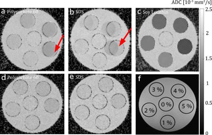
Fig. 7.
ADC maps of aqueous emulsifier solutions. The solutions were stored in CELLSTAR tubes and fixed in a cylindrical MR phantom. A central slice through the phantom is shown. (a–c) ADC maps obtained without additional inversion recovery preparation to suppress signal components caused by the emulsifiers (a: polysorbate, b: SDS, c: lecithin). Red arrows indicate chemical shift artifacts caused by the emulsifiers. (d–e) ADC maps acquired using an additional inversion recovery preparation for suppression of signal contributions from the emulsifiers themselves (d: polysorbate, e: SDS). (f) The arrangement of the samples with different emulsifier concentration in the MR phantom is depicted

