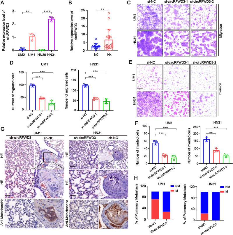
Fig. 2. CircRFWD3 could enhance the migration and invasion ability of HNSCC cells in vitro and in vivo.
A qRT-PCR assay showed the expression level of circRFWD3 in different HNSCC cell lines (UM1, UM2, HN30, and HN31). B A clinical cohort of nonmetastatic and metastatic HNSCC patient samples indicated significantly high expression of circRFWD3 in metastatic HNSCC patients (N0 = no lymph node metastasis, n = 15; Nx lymph node metastasis, n = 15). C, D The migration ability of UM1 and HN31 cells with silenced circRFWD3 was examined by transwell assays, scale bar, 100 μm. E, F The invasion ability of UM1 and HN31 cells by silencing circRFWD3 was examined by transwell assays, scale bar, 100 μm. G H&E and IHC analysis determined the metastatic tumor nodules in the lungs of mice injected with sh-NC or sh-circRFWD3 stably transfected UM1 and HN31 cells, scale bar, upper 100 μm, middle 10 μm. H Statistical graphs showed that the metastatic rate of the UM1 sh-NC group was 72.3% (n = 11), UM1 sh-cirCRFWD3 group was 27.3% (n = 11), HN31 sh-NC group was 25% (n = 12), and HN31 sh-cirCRFWD3 group was 0% (n = 12). NM no pulmonary metastasis, M pulmonary metastasis; Scale bar, 100 μm. Data were shown as the mean value ± SD of three independent experiments. The asterisks indicate significant differences (Student’s t-tests, *P < 0.05, **P < 0.01, ***P < 0.001).

