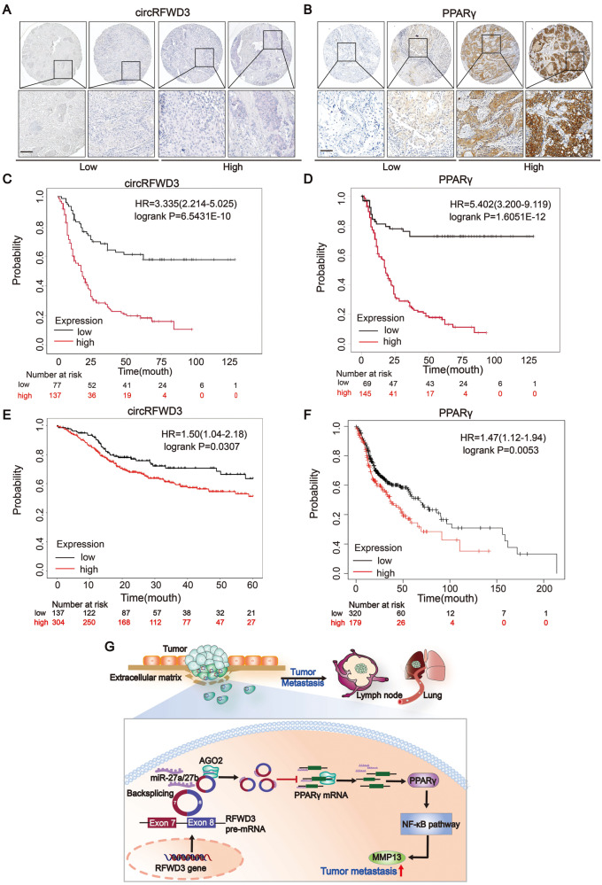
Fig. 7. The expression of circRFWD3 and PPARγ were negatively associated with prognosis in patients with HNSCC.
A, B ISH staining of circRFWD3(pink stain in the cytoplasm) and IHC staining of PPARγ in an HNSCC clinical cohort, scale bar, 10 μm. C, D Kaplan–Meier analysis showed that circRFWD3 and PPARγ were negatively correlated with the overall survival rate of HNSCC patients according to our clinical cohort. E, F Kaplan–Meier analysis showed that circRFWD3 and the mRNA expression of PPARγ were negatively correlated with the overall survival rate of HNSCC patients in the TCGA database. G Molecular mechanism of circRFWD3 involved in HNSCC metastasis. CircRFWD3, resulting from back-splicing of exons 7 and 8, could sponge-like bind to miR-27a/b to relieve the inhibitory effect of miR-27a/b on PPARγ, promoting the transcription and translation of PPARγ. Then, upregulated PPARγ could activate the NF-κB signaling pathway to promote the expression of MMP13 and accelerate tumor metastasis.

