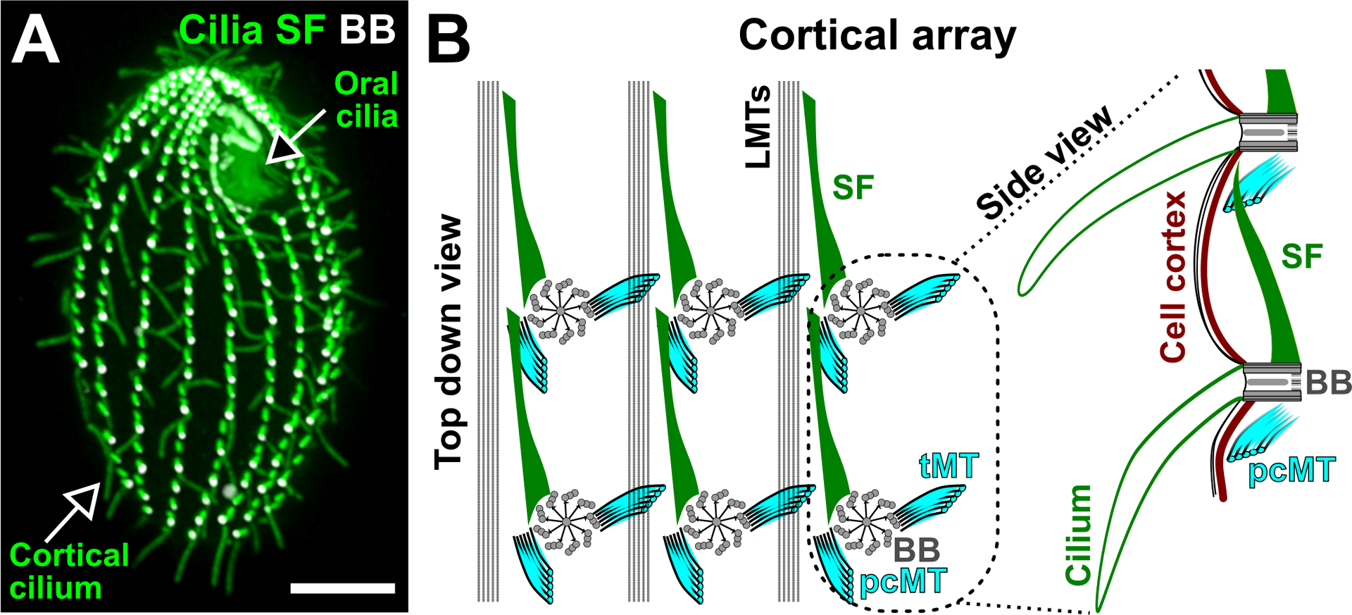
Figure 1.

Ciliate cortical architecture. (A) Image illustrates a Tetrahymena thermophila cell. The oral cilia are organized as a cluster at the cell anterior pole to promote feeding. The cortical cilia are arranged into longitudinal rows that are parallel to the cell’s AP axis to promote cell motility. Cilia and SF (green). BB (white). Bar, 10 μm. (B) left: Schematic depicting the top down view of the Tetrahymena cortical array. SFs (green) are oriented anteriorly, pcMTs (cyan) are oriented posteriorly and tMTs (cyan) are projected laterally. LMTs (black) span along the cell’s AP axis on the left of each BB (grey) row. BBs are connected by the BB-associated SF to the pcMT of the BB directly anterior to it. right: Schematic depicting the side view of two connected BBs and their associated cilia (green). The SF distal tip approaches the cell cortex (brown). Models are adapted from Soh et al. (2020).
