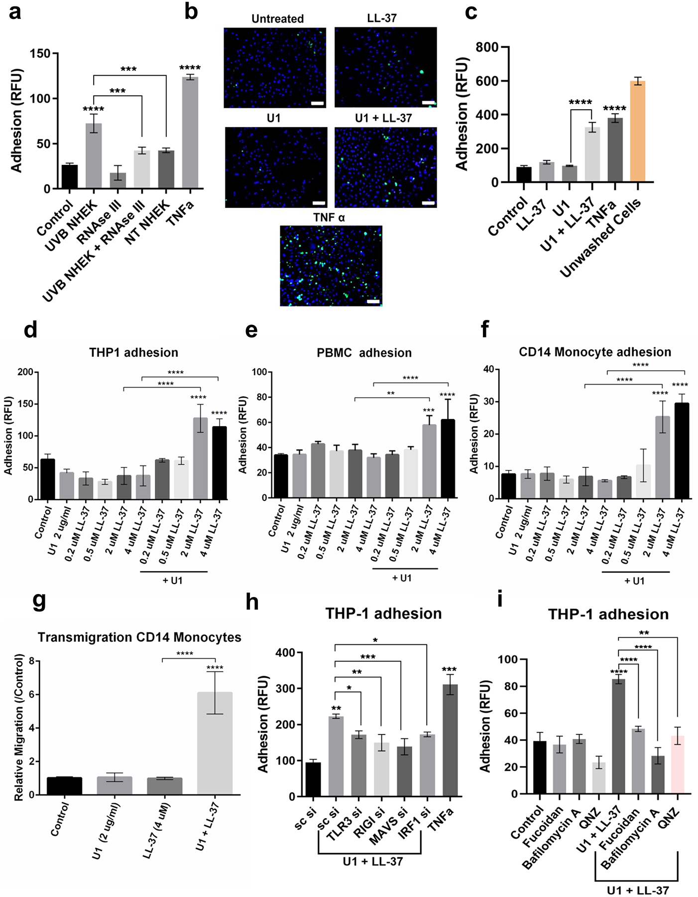
Figure 4: LL-37 and dsRNA enhance adhesion and migration of leukocytes across HDMECs.

(a) Non-treated and RNAse III treated UVB NHEK extracts added onto HDMECs for 12 hours and adhesion assay was performed with Calcein labeled THP-1 cells. (n=3). (b) Adhesion assay with THP-1 cells (green) was performed for HDMECs treated with U1 and/or LL-37 for 24 h. TNFα is positive control. DAPI (blue). Scale=50 μM. (c) Quantification of (b). (d, e, f) HDMECs treated with U1 (2 μg/ml) and/or LL-37 (0.2 μM-4 μM) for 24 h. Adhesion of labeled THP-1 monocytes (d), PBMCs (e) and CD14+ primary monocytes (f) to HDMECs was assessed. (n=3) (g) Trans-endothelial migration of Calcein labeled CD14+ primary monocytes (n=3) (h) siRNAs against TLR3, RIGI, MAVS and IRF1 in HDMECs, followed by treatment with U1 and LL-37. Adhesion assay with labeled THP-1 monocytes (n=3). (i) THP1 adhesion assay for HDMECs pre-treated with fucoidan (50 μg/ml), Bafilomycin A1 (100 nM) and QNZ (100 nM) for 60 min, followed by U1 and LL-37 treatment for 24 h. (*, p< 0.05; **, p<0.01; ***, p<0.001; ****, p<0.0001)
