
FIGURE 2.
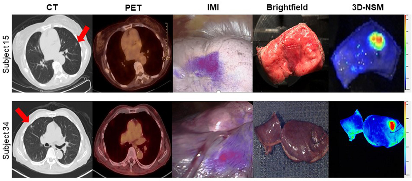
Three-dimensional near-infrared specimen mapping (3D-NSM) localizes nonpalpable pulmonary lesions. This figure shows representative images from 2 patients with nonpalpable pulmonary lesions enrolled in the study. Preoperative computed tomography (CT) and positron emission tomography (PET) images are shown on the left with red arrows denoting lesions. The center panel shows brightfield images in which lesions cannot clearly be identified by visual inspection and palpation but are highly fluorescent on 3D-NSM.