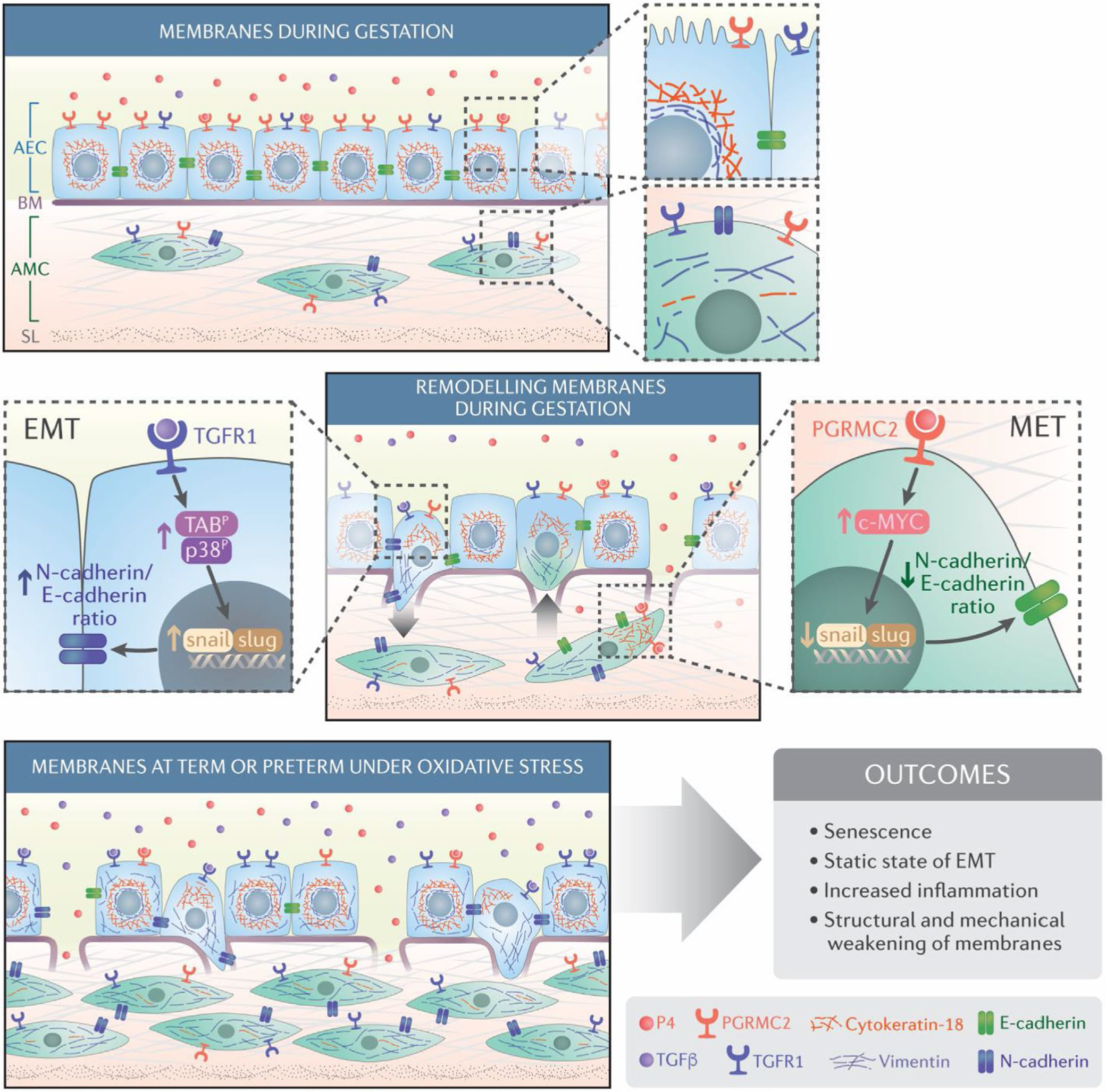
FIGURE 2.

Schematic representation of maintenance of the fetal membrane integrity and its disruption due to cellular transitions: The Amnion membrane is comprised of amnion epithelial cells (AEC) and amnion mesenchymal cells (AMC) separated by a Type 4 basement membrane collagen (BM) and extracellular matrix (ECM). During gestation, progesterone (P4) helps to maintain AEC epithelial state. AMCs, located in the extracellular matrix, express a fibroblastoid morphology. The ratio between AEC and AMC is 10:1 in normal gestation. EMT is mediated by TGF-β/TGF-β receptor-mediated mechanisms regulated by p38MAPK (middle panel; left). P4 and progesterone membrane receptor component (PGRMC2) mediated MET recycles AMC back to AEC to maintain tissue homeostasis and prevent accumulation of AMCs in the membrane extracellular matrix p38MAPK (middle panel; right). Oxidative stress causes increased activation of p38MAPK at term or preterm leading to increased EMT and a reduction in the function of progesterone as oxidative stress down-regulates PGRMC2 in AMCs. As the recycling of cells is stalled, AMC accumulates in the ECM and causes inflammatory changes (bottom panel). This figure was originally publsihed in Sci Signaling Richardson et al.173 reproduced here with permission
