
Figure 2. Multiplex two-photon imaging with miRFP670nano3 in the brain and spinal cord of live mice.
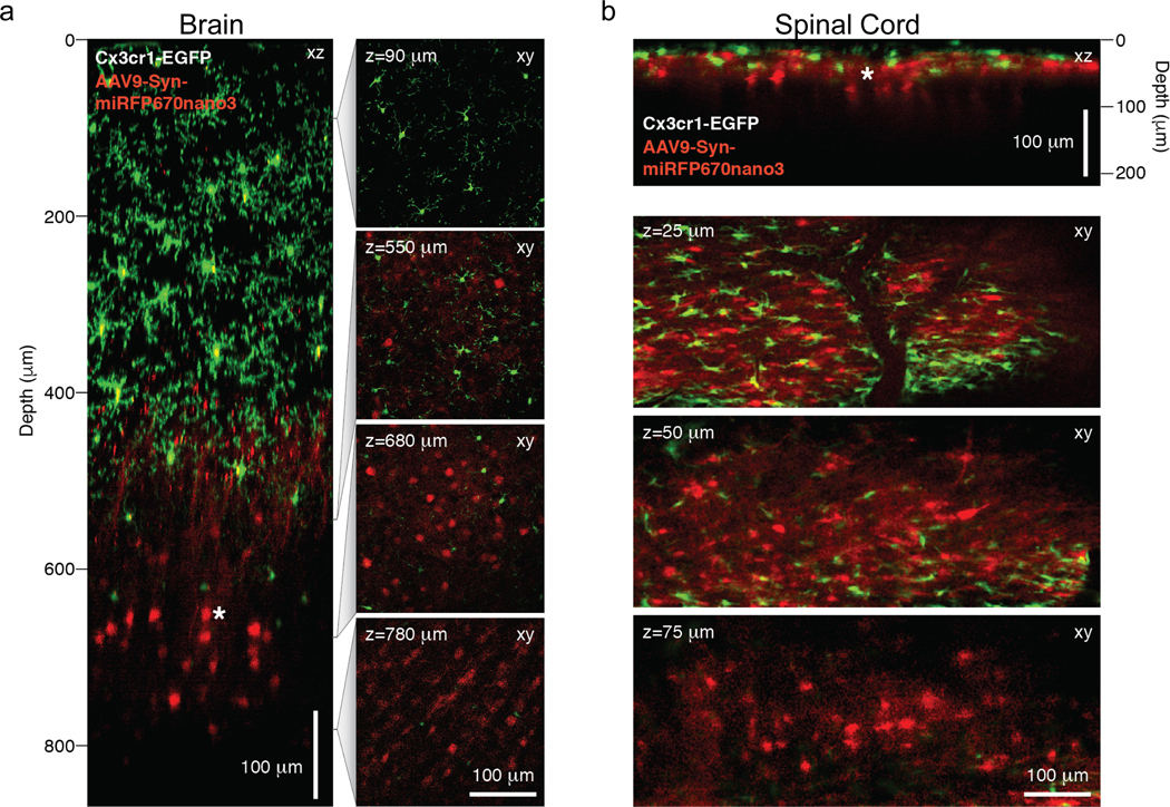
(a) Left, xz sub-projection from a xy fluorescence image stack showing neurons (red) and microglia (green) in the somatosensory cortex of a 14-weeks-old Cx3cr1 GFP/+ mouse five weeks after stereotactic AAV9-hSYN-miRFP670nano3 vector delivery into deep cortical layers. Right, four example images from the xy fluorescence image stack at the indicated depths (z) from the pial surface. (b) Top, xz sub-projection from a xy fluorescence image stack showing neurons (red) and microglia (green) in the lumbar spinal cord of a 12.5-weeks-old Cx3cr1 GFP/+ mouse five weeks after stereotactic AAV9-hSYN-miRFP670nano3 vector delivery into superficial dorsal horn laminae. Bottom, three example images from the xy fluorescence image stack at the indicated depths (z) from the pial surface. (a-b) GFP and miRFP670nano3 fluorescence images were acquired simultaneously using a 920 nm excitation light and emission filters 525/70 nm for EGFP and 645/75 nm for miRFP670nano3. No external BV was administered. Representative images of three experiments are shown. Scale bars, 100 μm.