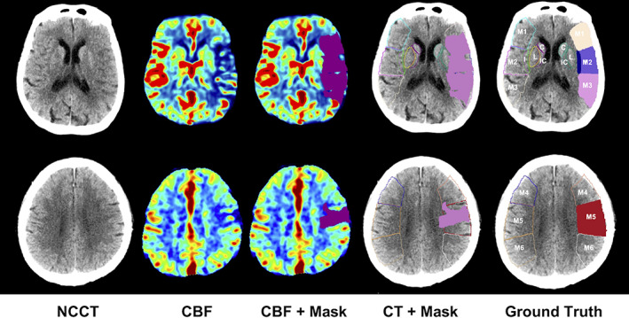
FIGURE 2.

Procedure to extract the ischemic cores from CBF and determine the affected ASPECTS regions in CT. The rows are for two image slices of a representative patient. The first column shows the input NCCT image slices. The second column shows the related CBF images. The third column shows the ischemic core defined on CBF with the rule of rCBF <30%. The fourth column matches the core region on NCCT. In the last column, an ASPECTS region is defined as affected if the ratio of the core volume in this region over the region volume
