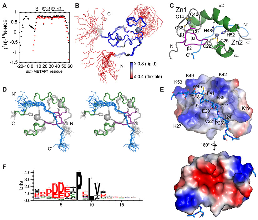
Figure 3. NMR structure of METAP1-ZNG1 complex.
(A) 1H,15N NOE for the fusion (black) and free domain (red). (B) Structure of the 20 lowest-energy conformers of the Mm ZNG110-30 METAP11-59 fusion, colored by 1H,15N NOE. (C) Ribbon diagram of Mm ZNG110-30 METAP11-59 showing Zn coordination. (D) Stereo view of 20 lowest-energy conformers of Mm ZNG110-30 METAP11-59, colored by secondary structure, with helices in green, beta strands in purple, and peptide in blue. (E) Electrostatic surface map of the METAP11-59 peptide interface. (F) N-terminal ZNG1 motif among eukaryotic cluster 3 COG0523 proteins. See also Figure S3 and Table S2.

