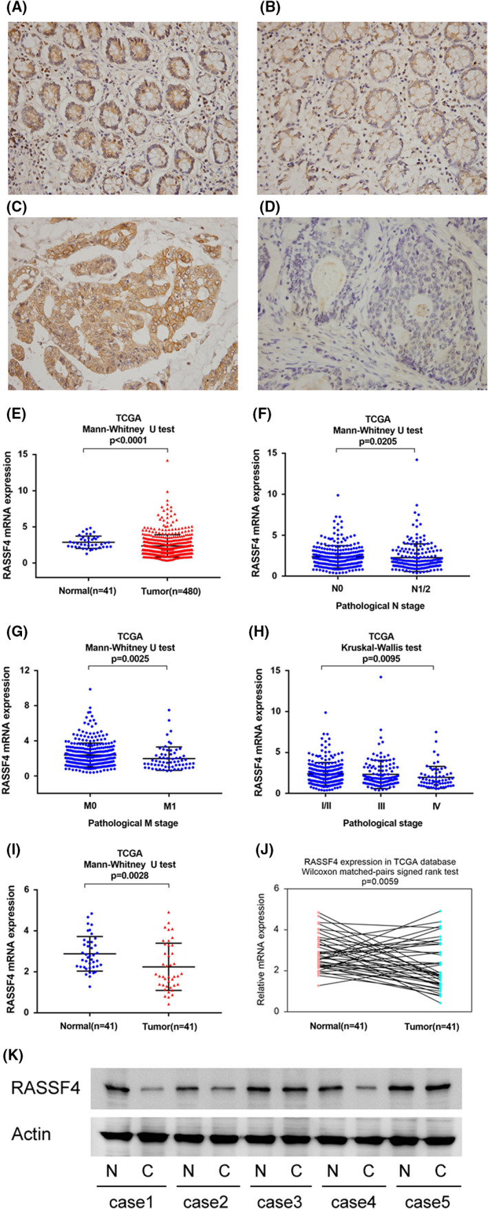
FIGURE 1.

Expression of RASSF4 in colorectal cancer (CRC). (A) Strong RASSF4 expression in normal colon tissue. (B) Moderate RASSF4 expression in a case of normal colon tissue. (C) Strong cytoplasmic RASSF4 expression in a case of CRC. (D) Negative RASSF4 expression in a case of CRC. (E) The Cancer Genome Atlas (TCGA) colorectal cancer cohort analysis. Expression of RASSF4 in CRC tissues was significantly lower than that in normal colon tissues (p < 0.0001). (F) TCGA analysis showed that RASSF4 expression was lower in CRC tissues with positive nodal metastasis (p = 0.0205). (G) TCGA analysis showed that RASSF4 expression was lower in colorectal cancers with distal metastasis (p = 0.0025). (H) TCGA analysis showed that RASSF4 expression was significantly lower in colorectal cancers with higher pathological stage (p = 0.0095). (I and J) Analysis of 41 cases of paired tumour/normal samples from TCGA showed that RASSF4 was lower in CRC tissues compared with corresponding adjacent normal colon tissues (p < 0.05). (K) Western blot in five colorectal cancer tissues and paired adjacent normal tissues showed RASSF4 downregulation in two out of five of paired cases
