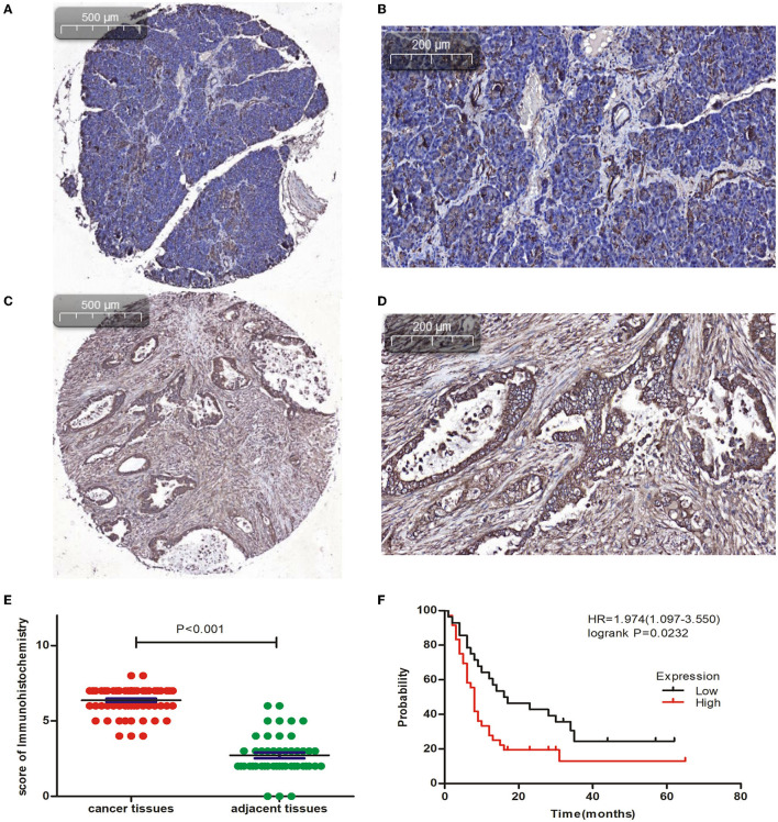
Figure 4.
Immunohistochemical analysis of CAPN2 expression in human PC tissues. (A,B) Representative IHC staining images of CAPN2 protein in noncancerous pancreatic tissues and (C,D) PC tissues at X5 and X20 magnification, respectively. (E) Comparison of the CAPN2 expression score shows that CAPN2 protein levels are significantly higher (P < 0.001) in PC tissues (N = 64) than in noncancerous pancreatic tissues (N = 54). (F) Kaplan–Meier analysis shows that high expression of CAPN2 is associated with poorer OS than low expression of CAPN2 in patients with PC (HR = 1.974, P = 0.0232).

