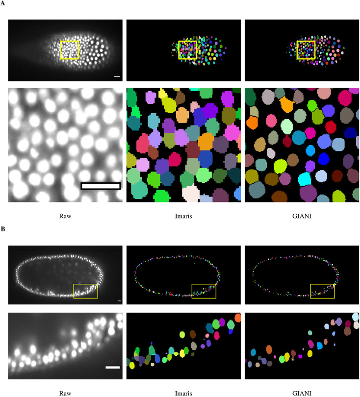
Fig. 6.
Demonstration of GIANI on a large light-sheet microscopy dataset. Illustrations of the segmentations produced by GIANI and Imaris on a Tribolium castaneum embryo dataset derived from light-sheet microscopy (available to download from https://doi.org/10.5281/zenodo.5270323). Two different slices of the 3D volumes are shown, at ∼46 μm (A) and ∼247 μm (B), to illustrate the variation in nuclei morphologies at different depths. In each case, the top row shows the relevant slice of each 3D volume, whereas the bottom row shows the magnified views of the boxes in the top row images. ∼5600 nuclei were detected in the full volume. Scale bars: 20 μm. The GIANI and Imaris settings used to produce this data are available to download from https://doi.org/10.5281/zenodo.6206042.

