FIGURE 1.

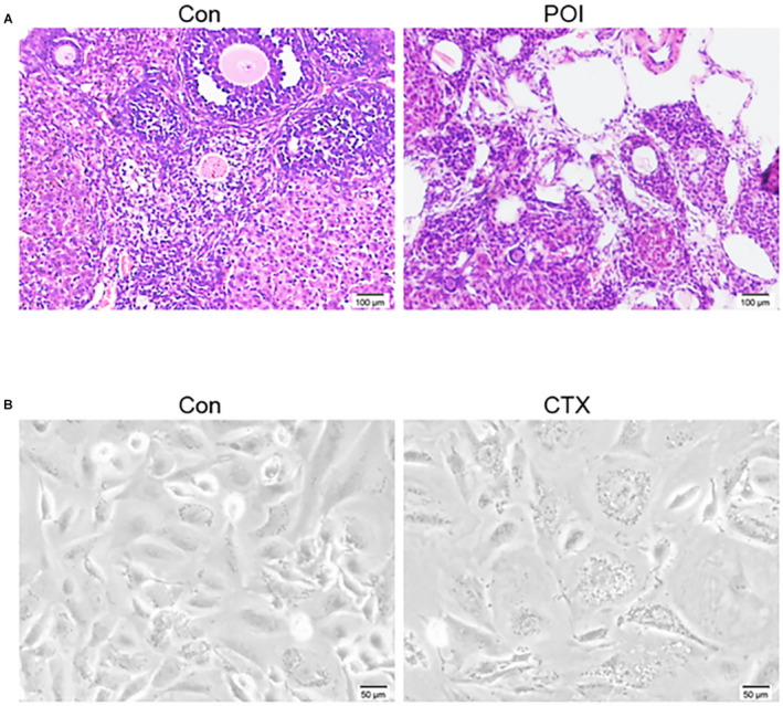
Typical characteristics of the ovary and granulosa cells (GCs) of normal and mice treated with chemotherapy drugs. (A) Histological analysis of ovarian sections of 6 weeks old mice after 2 weeks of treatment with (right) or without (left) cyclophosphamide (CTX) and busulfan, a representative hematoxylin and eosin staining of 15 mouse ovaries. Scale bar: 100 μm; (B) A representative GCs morphology of normal (left) or with CTX treatment (right), the cells were polygonal and fiber‐like; compared with the control group, the number of ovarian GCs in the CTX group was significantly reduced. Scale bar: 50 μm
