FIG. 2.
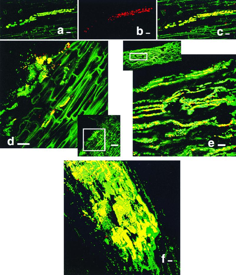
Immunolocalization of NifH produced by K. pneumoniae 2028 in maize roots. All images are longitudinal sections. (a to c) Series of images demonstrating fluorescence generated by green GFP-labeled cells (a), red NifH-producing cells (b), and a combination of the preceding images resulting in yellow cells (c). Immunolocalization of NifH at 3 (d), 4 (e), and 8 (f) days after inoculation. Bar, 10 (a to c), 20 (d, e, and f), or 50 (d offset) μm.

